18 March 2021: Animal Study
Functional Dissection of CD26 and Its Pharmacological Inhibition by Sitagliptin During Skin Wound Healing
Yue Jiang 1BCD* , Yuan Yao 1BCF* , Jin Li 1AF , Yanling Wang 1CF , Jie Cheng 2DE , Yumin Zhu 1ADEG*DOI: 10.12659/MSM.928933
Med Sci Monit 2021; 27:e928933
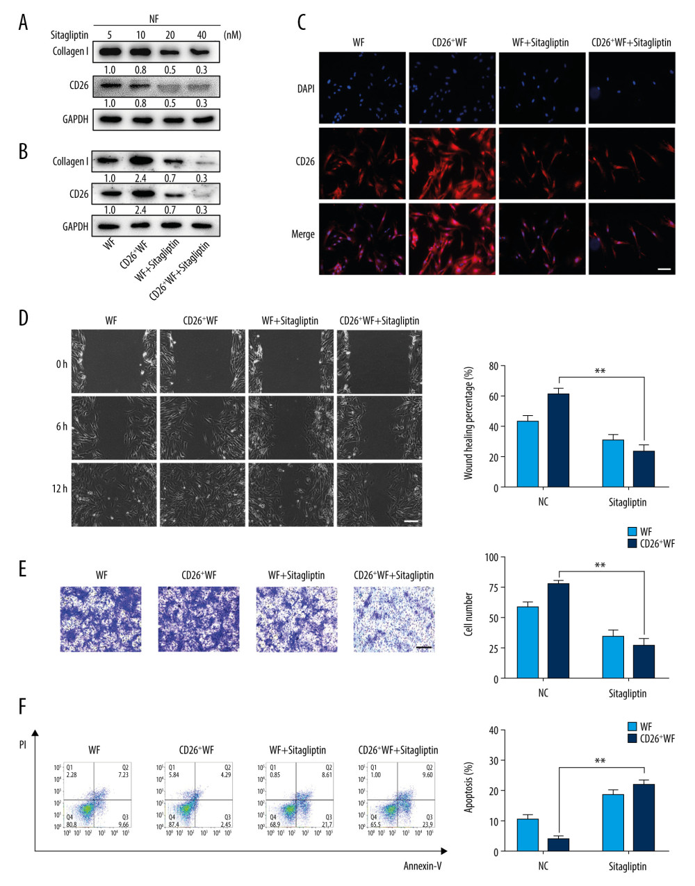
Figure 3 Sitagliptin inhibits the pathological features of CD26+ wound fibroblasts in vitro. (A) The expression of CD26 and Col1 in WFs and CD26+ WFs was measured by western blot following a concentration gradient of sitagliptin treatment. Representative images are shown. (B) Immunofluorescent staining of CD26 in WFs and CD26+ WFs after sitagliptin treatment. Representative images are shown. (C) The migration ability of CD26+ WFs was significantly reduced following sitagliptin treatment (20 nM) in wound healing assays. (D) The migration ability of CD26+ WFs was significantly reduced after sitagliptin treatment (20 nM) in transwell assays. (E, F) An evident increase in apoptosis within the WFs and CD26+ WFs following sitagliptin treatment (20 nM) as assayed by Annexin V-PI staining and its quantification (n=5). Scale bar: 100 μm. t test, * P<0.05 vs WF group, ** P<0.01 vs WF group.