10 March 2021: Clinical Research
High-Resolution Magnetic Resonance Imaging (HR-MRI) Imaging Characteristics of Vertebral Artery Dissection with Negative MR Routine Scan and Hypoperfusion in Arterial Spin Labeling
Yonggang Zhang BCDEG* , Chongchang Miao CDEG* , Yan Gu ACDF* , Shunbin Jiang BD , Jian Xu ABCDOI: 10.12659/MSM.929445
Med Sci Monit 2021; 27:e929445
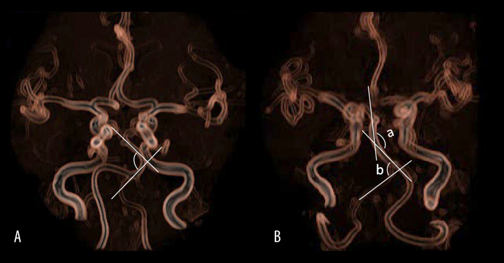
Figure 1 (A) is a schematic diagram to measure the bending angle of the basilar artery. The arrow indicates the angle formed by 2 sides of the basilar artery curvature toward the midline. (B) is a schematic diagram to measure the vertebrobasilar artery minimum angle. Take the smallest angle of the distal base artery or the V4 segment of the vertebral artery in the dissection lesion. The vertebral-basal artery at the distal end of the dissection lesion formed 2 angles, which were the basilar artery bending angle and the vertebral artery bending angle. The vertebral artery angle was 85°, while the basilar artery bending angle was 135°. Therefore, the vertebrobasilar artery minimum angle was 85°.