16 June 2021: Review Articles
Current Applications and Selected Technical Details of Dual-Energy X-Ray Absorptiometry
Piotr Sawicki 1ABCDEF* , Marek Tałałaj 2D , Katarzyna Życińska 13DEG , Wojciech S. Zgliczyński 4DFG , Waldemar Wierzba 56DEFGDOI: 10.12659/MSM.930839
Med Sci Monit 2021; 27:e930839
Background
Importance of the DXA in Diagnostics of BMD
Safety Considerations Regarding Radiation Dose
DXA Diagnosis of Vertebral Fractures Using VFA
Visualization of Abdominal Aortic Calcifications (AAC)
Evaluation of Hip Structure Analysis (HSA)
Diagnosis of Atypical Femur Fracture (AFF)
Evaluation of Finite Element Analysis (FEA)
Evaluation of FRAX
Definition and Importance of TBS
Body Composition Assessment
Evaluation of Periprosthetic DXA
Other Functions
Conclusions
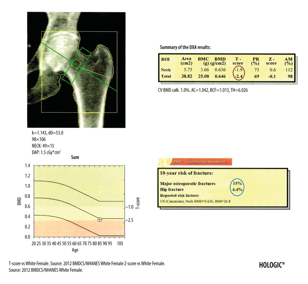
Figure 2 DXA examination of the hip. The diagnostic result is the T-score parameter estimated for the femoral neck (the upper value in the red circle) and for the total proximal femur (the lower value in the red circle). The ROI of the femoral neck (marked as green frame) should be perpendicular to the femoral neck, should not include the greater trochanter and the ischial bone, but should include soft tissues on both sides of the femoral neck. The midline (marked as green line) should cover the long axis of the femoral neck. In the blue circle is marked the FRAX for major osteoporotic fracture (upper value) and FRAX for hip fracture (lower value).