31 March 2024: Review Articles
Differentiation of Native Vertebral Osteomyelitis: A Comprehensive Review of Imaging Techniques and Future Applications
Weijian Zhu 12BCEF , Sirui Zhou 3D , Jinming Zhang 1D , Li Li 4B , Pin Liu 2A , Wei Xiong 1A*DOI: 10.12659/MSM.943168
Med Sci Monit 2024; 30:e943168
Introduction
Segmental Structure of the Spine
Diagnostic Value of Plain Radiographs and Computed Tomography for NVO
Diagnostic Value of MRI in NVO
Pyogenic Spondylitis
Tuberculous Spondylitis
Brucellar Spondylitis
Fungal Spondylitis
Diagnostic Value of Positron Emission Tomography for NVO
Artificial Intelligence Applications and the Future
Conclusions
References
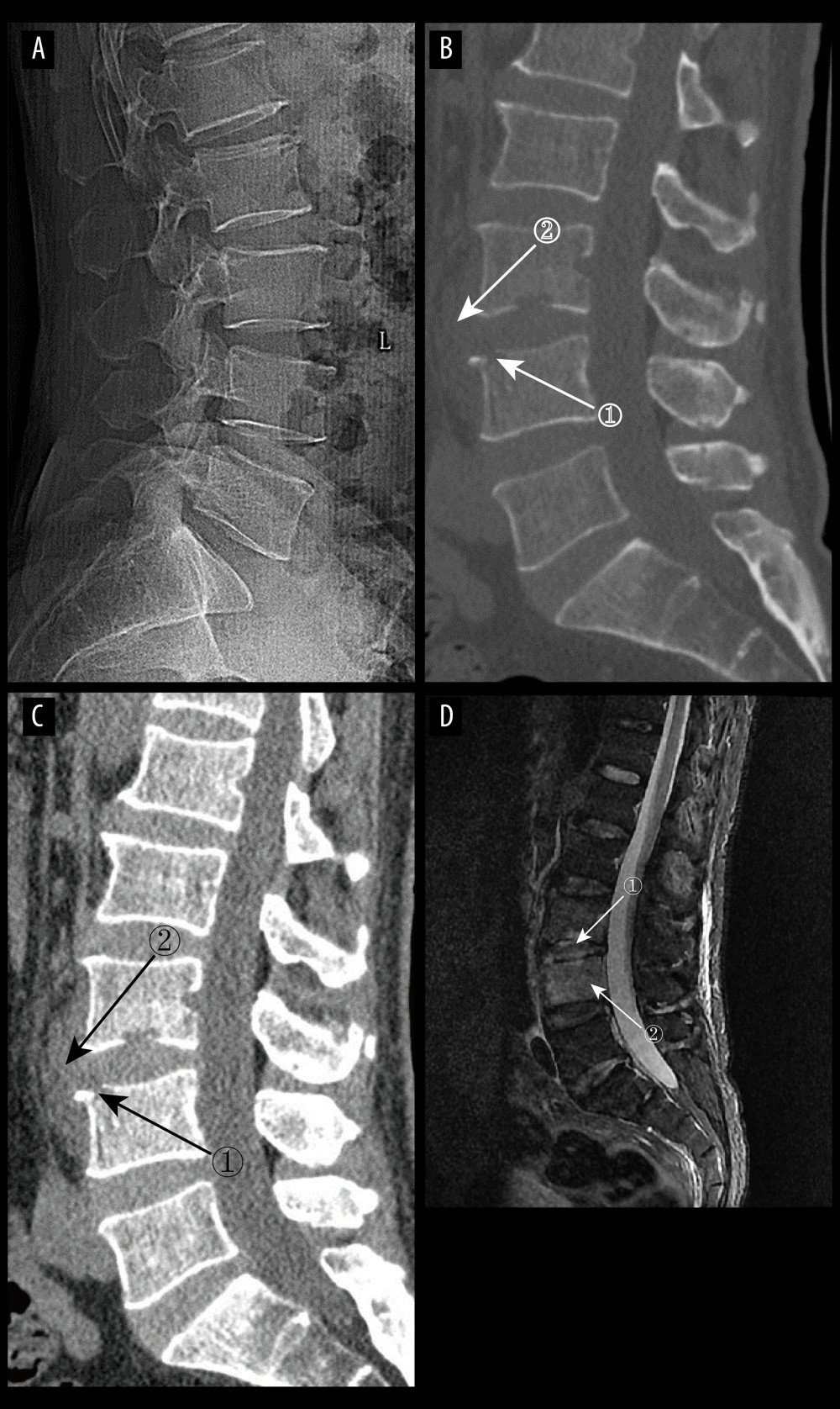
Figure 3 (A) pyogenic spondylitis (PS) early plain radiograph. No significant narrowing of the lumbar spinal space was seen. (A–D) Images of the same patient, who presented with 9 days of low back pain. (B, C) Plain and enhanced computed tomography in early PS. Marker 1 in both images shows poorly defined bony margins on the anterosuperior margin of the L4 vertebral body, and marker 2 shows swelling of the surrounding soft tissues (D) Magnetic resonance imaging in early stages of PS. Marker 1 shows disc erosion, and marker 2 shows diffuse infection of the L4 vertebral body (Adobe Illustrator 2022. 26.5. Adobe Inc.).