31 March 2024: Review Articles
Differentiation of Native Vertebral Osteomyelitis: A Comprehensive Review of Imaging Techniques and Future Applications
Weijian Zhu 12BCEF , Sirui Zhou 3D , Jinming Zhang 1D , Li Li 4B , Pin Liu 2A , Wei Xiong 1A*DOI: 10.12659/MSM.943168
Med Sci Monit 2024; 30:e943168
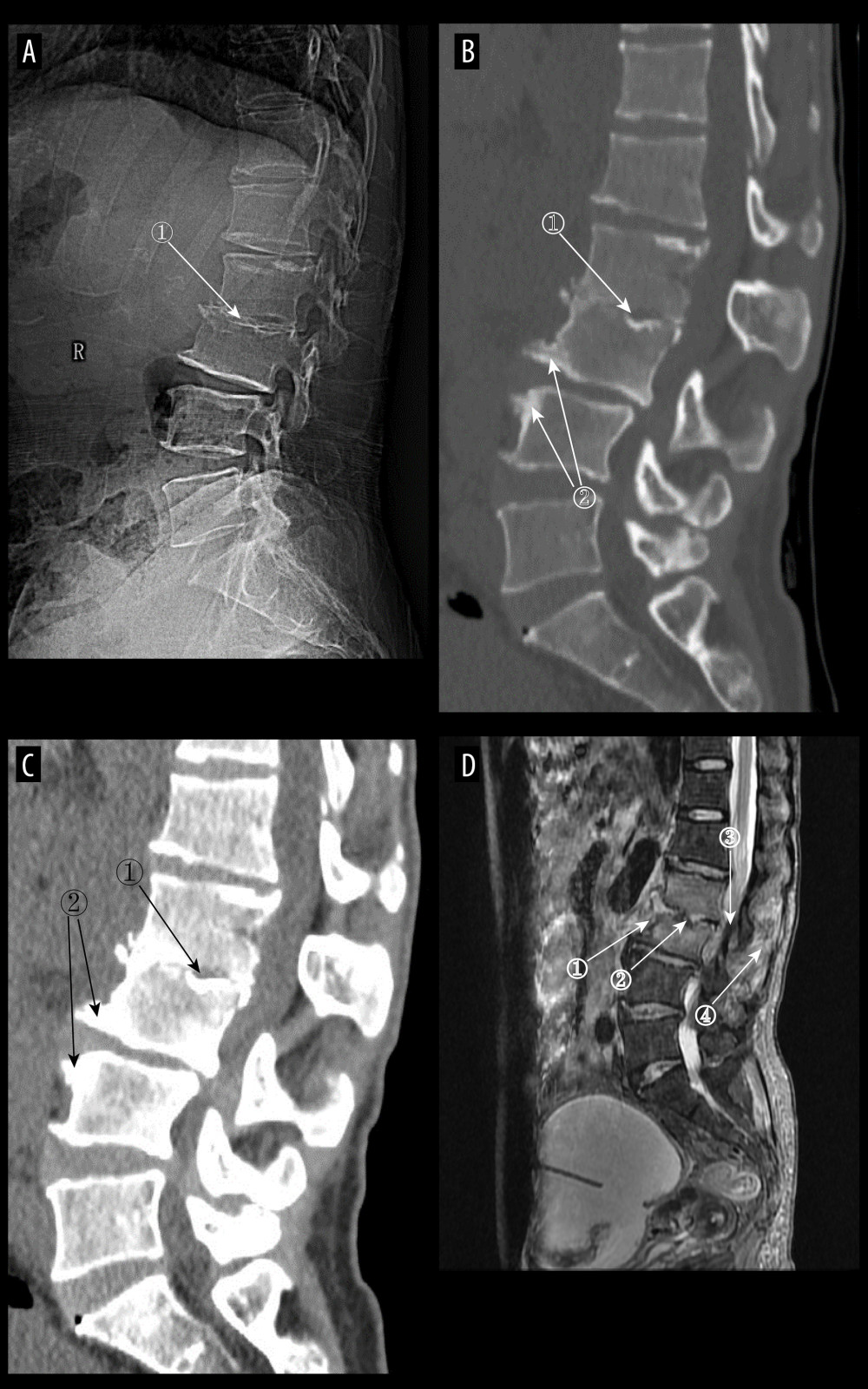
Figure 4 (A) Plain radiographs of late pyogenic spondylitis (PS). Marker 1 shows severe narrowing of the L2–3 intervertebral space, with bony hyperplasia and sharpening of the vertebral margins. (A–D) Images of the same patient, with 42 days between onset of symptoms and the time of the radiograph. (B, C) Plain and enhanced computed tomography in late-stage PS. Marker 1 shows marked narrowing of the L2–3 intervertebral space, and marker 2 shows rough and blurred edges of the L3–L4 vertebral body with osteolytic changes. (D) Magnetic resonance imaging of late PS. Osteolysis of the anterior margin of L3 is shown at marker 1, severe narrowing of the L2–3 intervertebral space is shown at marker 2, osteophytes of the small joints are shown at marker 3, and high signal of the lumbar attachments is shown at marker 4 (Adobe Illustrator 2022. 26.5. Adobe Inc.).