31 March 2024: Review Articles
Differentiation of Native Vertebral Osteomyelitis: A Comprehensive Review of Imaging Techniques and Future Applications
Weijian Zhu 12BCEF , Sirui Zhou 3D , Jinming Zhang 1D , Li Li 4B , Pin Liu 2A , Wei Xiong 1A*DOI: 10.12659/MSM.943168
Med Sci Monit 2024; 30:e943168
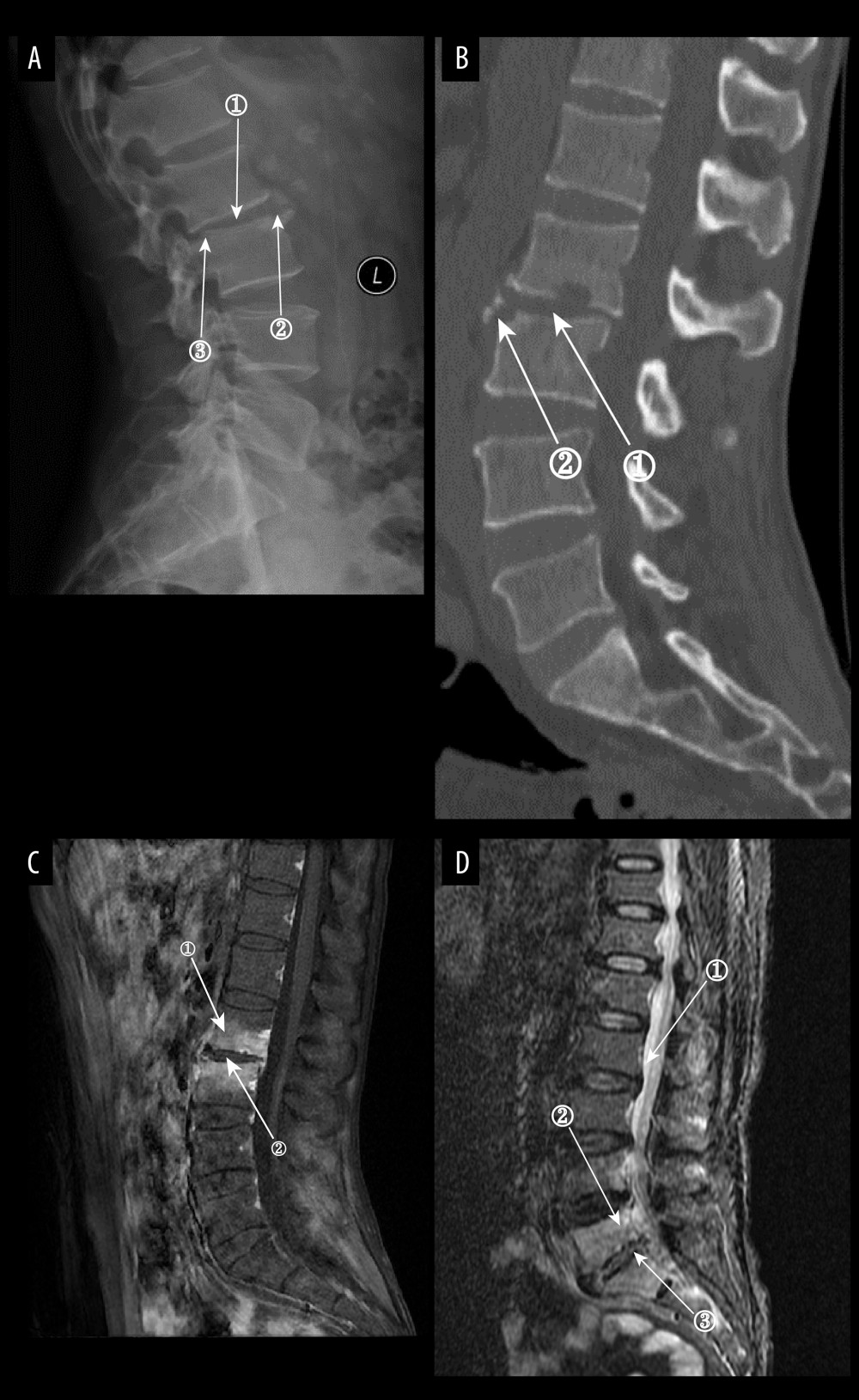
Figure 7 (A) Plain radiographs of late Borrelia burgdorferi spondylitis (BS). Marker 1 shows a decrease in the height of the intervertebral space, marker 2 shows osteophytes on the margins of the vertebral body, and marker 3 shows a slight forward displacement of the L3 vertebral body. (A–D) Images of the same patient, with an interval of approximately 120 days between the onset of symptoms and the time of the radiograph. (B) Plain computed tomography in late BS. In both figures, Marker 1 shows a decrease in the height of the intervertebral space, and marker 2 shows hyperplasia of the anterior margin of the vertebrae. (C) Magnetic resonance imaging (MRI) of late BS. This image is a T1-enhanced image showing abnormal signal in the vertebral body at marker 1 and reduced intervertebral space height in the vertebral body at marker. (D) MRI of late BS. Density at marker 1 is 54, marker 2 shows diffuse infection of the vertebral body with a density of 37, and marker 3 shows an infected disc. The time between the onset of this patient’s disease and the radiograph is about 2 years. (ITK-SNAP. Version 4.0.2. Paul Yushkevich, Jilei Hao, Alison Pouch, Sadhana Ravikumar et al at the Penn Image Computing and Science Laboratory; Adobe Illustrator 2022. 26.5. Adobe Inc.).